
Electronic Enhancement
Video Enhancement
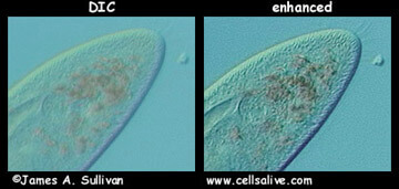
Once contrast in the microscope image is optimized, the image can be captured and further enhanced using a video camera. These cameras allow integration (amplification) of low light signals and stepwise adjustment of contrast and brightness of the integrated signal. Video contrast enhancement is illustrated in this Entamoeba histolytica imaged by DIC (right).
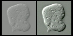
Increasing Apparent Resolution
Some video cameras used for research also have the capability of stepwise adjustment of "sharpness". Much like the sharpen filter in programs like Photoshop ®, this spatial filter enhances the apparent resolution of the image in real time. This is particularly useful in specimens that move quickly - the sharpness can be adjusted once and the enhanced specimen recorded. AParamecium (right) is seen in both unenhanced and enhanced DIC camera images.
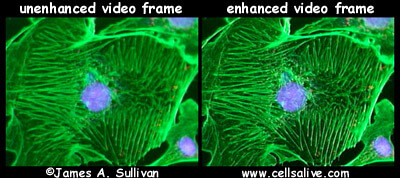
Fluorescence Enhanced